Cell-Based Phenotypic Screening
INQUIRYRelying on advanced automated screening platforms, mature cell model systems, and multidimensional high-content imaging and analysis technologies, we are capable of accurately evaluating the biological activity and mechanistic effects of compounds at the cellular level. Whether it is standard cytotoxicity screening, signal pathway regulation analysis, or customized evaluations targeting complex disease phenotypes, BOC Sciences offers a one-stop solution encompassing compound library construction, screening program design, experimental execution, and data interpretation. Our professional team has extensive project experience and can support clients in the early discovery and optimization of drugs with unknown targets or unclear mechanisms, significantly improving the efficiency of lead compound screening.
Service Highlights and Advantages
- Compound library screening or evaluation of customer-provided compounds: We offer screening of over 10,000 ready-to-use small molecules or evaluation of 1–1000 customer-provided compounds.
- High-throughput and high-content screening platform: Capable of screening over 10,000 compounds in a single run, supporting multi-channel HCS image acquisition and multi-parameter analysis.
- Rich cell model resources: Covering over 50 tumor, neural, and immune cell lines, with support for custom client-provided cells.
- Diverse phenotypic detection indicators: Offering more than 20 indicators, such as LC3/Caspase staining, autophagy flux detection, cell cycle distribution, etc.
- Seamless integration with mechanism studies: Downstream analysis options include proteomics, RNA-seq, Western blot, or fluorescence co-localization.
- Combination drug screening and synergy evaluation: Supporting 2×2 or 3×3 drug combination matrix analysis and synergy index calculation.
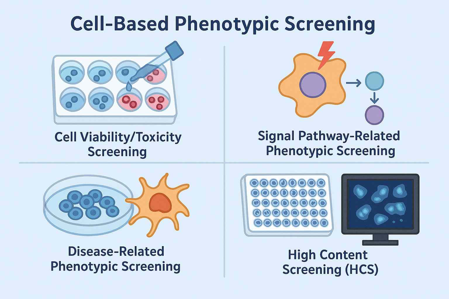
Cell-Based Phenotypic Screening Services by BOC Sciences
BOC Sciences has established a highly integrated, standardized, and customizable in vitro phenotypic screening platform supported by advanced cell culture facilities, automated high-throughput screening systems, high-content analysis platforms (HCA/HCS), and multi-mode detection technologies. Our services encompass the entire process from cell model construction, screening system optimization, compound library screening, image and data analysis to mechanism validation, fully meeting the diverse needs of clients in biopharmaceutical, biotechnology, and research institutions.
Cell Viability/Toxicity Screening
BOC Sciences offers a variety of detection methods based on metabolic activity, membrane integrity, and cell viability indicators. We provide a model library of various cancer cell lines, primary cells, and stem cells for screening, and can customize cell lines or labeling systems according to customer requirements.
- MTT/MTS/CCK-8 colorimetric assay.
- ATP fluorescence assay (CellTiter-Glo®).
- Annexin V/PI dual-staining flow cytometry.
- LDH release assay.
- Caspase activity analysis.
Signal Pathway-Related Phenotypic Screening
By combining high-content imaging technologies, we collect multidimensional data on organelle localization, nuclear translocation, phosphorylation status, and expression of upstream/downstream proteins in signaling pathways. Using fluorescence/luciferase reporter gene systems, antibody labeling, and immunofluorescence imaging, we can assess the activation or inhibition of key signaling pathways by small molecule compounds:
- NF-κB pathway.
- Wnt/β-catenin pathway.
- PI3K/Akt/mTOR pathway.
- MAPK/ERK pathway.
- p53/MDM2 pathway.
Disease-Related Phenotypic Screening
BOC Sciences specializes in constructing disease-specific cell models to simulate real pathological processes, enabling precise phenotypic screening. We support the use of CRISPR, RNAi, and other gene-editing tools to establish disease-relevant cell lines and can integrate co-culture systems or 3D cell models to enhance physiological relevance and predictive capability.
- Neurodegenerative disease models: Assessment of neurite outgrowth, axonal degeneration, mitochondrial function, Tau protein aggregation, etc.
- Metabolic disease models: Lipid droplet accumulation, insulin resistance-related signaling.
- Inflammatory models: Macrophage activation, inflammatory cytokine release (e.g., IL-6, TNF-α).
- Viral infection models: Viral replication, host antiviral response.
- Tumor metastasis models: Cell migration/invasion, epithelial-mesenchymal transition (EMT), etc.
High Content Screening (HCS)
HCS is one of BOC Sciences' core services. By integrating fluorescent staining, multi-channel high-resolution imaging, and image recognition algorithms, we perform quantitative analysis of cellular phenotypes across multiple parameters, indicators, and time points. Detectable phenotypes include but are not limited to:
- Changes in cell morphology and organelle localization.
- Cytoskeleton rearrangement (microtubules, actin).
- Nuclear translocation events (e.g., NF-κB, STAT1).
- Multi-target co-localization analysis.
- Multi-stain co-fluorescence evaluation.
- Neuronal phenotypes such as axonal length and branching point count.
Do You Need A Consultation?
BOC Sciences integrates innovative technologies to empower your drug discovery with strong momentum, fully dedicated to building next-generation drug screening platforms.
Phenotypic Indicators Supported by BOC Sciences
In in vitro phenotypic screening, the range of detectable cellular behaviors or functional indicators is extremely broad, covering multiple dimensions such as cell growth, differentiation, signal transduction, metabolic changes, and structural remodeling. Leveraging high-throughput imaging, high-content analysis, flow cytometry, and multiplex fluorescent labeling, BOC Sciences can efficiently quantify the following representative phenotypic features:
| Phenotypic Category | Functional Description | Common Detection Methods / Markers |
|---|---|---|
| Cell Proliferation & Death | Evaluates whether compounds promote or inhibit cell growth, and assesses cytotoxic or protective effects. | MTT/MTS, CCK-8, ATP-based luminescence, LDH release assay, cell counting, live/dead cell staining. |
| Autophagy & Apoptosis | Monitors programmed cell death, critical for studies in oncology and neurodegenerative diseases. | LC3, Beclin-1, Caspase-3/7 activity, Annexin V/PI staining, TUNEL assay. |
| Cell Cycle Analysis | Determines whether compounds induce cell cycle arrest at G1, S, or G2/M phases. | PI staining with flow cytometry, BrdU/EdU incorporation assays. |
| Cell Migration & Invasion | Assesses the ability of tumor or immune cells to migrate and invade, relevant for anti-metastasis research. | Wound healing assay, Transwell migration/invasion assays, time-lapse microscopy. |
| Cell Secretion Function | Measures secretion of cytokines, neurotransmitters, hormones, antibodies, etc. | ELISA, Luminex, multiplex assays, Western blot, supernatant analysis. |
| Organelle Morphology & Function | Evaluates drug-induced changes in organelle function, important for toxicity and mechanism studies. | JC-1 for mitochondrial membrane potential, ROS detection, ER stress markers (CHOP, GRP78), Lysotracker. |
| Receptor Internalization & Nuclear Translocation | Observes internalization of membrane receptors (e.g., GPCRs) or nuclear translocation of transcription factors (e.g., NF-κB, STAT). | Fluorescent protein labeling, immunofluorescence, nuclear-cytoplasmic fractionation with Western blot, reporter assays. |
| Axon Growth & Synapse Formation | Applied in neuronal development, regeneration, and plasticity research, essential for CNS drug screening. | Neuron-specific markers (βIII-tubulin), synaptic markers (Synapsin, PSD-95), image-based quantification. |
| Cytoskeletal Rearrangement | Evaluates the impact of compounds on cytoskeletal structures such as microtubules and actin filaments. | Phalloidin for F-actin staining, α-tubulin antibodies, confocal microscopy. |
| Cell Stress & Metabolic Status | Assesses oxidative stress, metabolic disruption, and related functional states. | ROS detection, NAD⁺/NADH ratio, AMPK activity, glucose uptake/lactate production assays. |
Project Workflow

Project Requirement Communication and Solution Design
After clients provide screening targets and specific requirements, our scientific team conducts a comprehensive project evaluation. This includes screening objectives (e.g., anti-tumor, neuroprotection), expected phenotypes (e.g., apoptosis, autophagy), selection of cell models, screening methodology, and endpoint detection indicators. A complete experimental proposal will be developed and executed upon mutual confirmation.
Cell Model Construction and Preliminary Experiment
Based on client requirements, we select standard cell lines (or use client-provided cells) to construct appropriate models. For disease models, stable transfected cell lines, or reporter gene constructs, personalized customization is also available. During the preliminary experiment phase, we optimize culture conditions, stimulation protocols, treatment concentrations, and endpoint detection methods to ensure data reliability and reproducibility.
Compound Treatment and Screening Execution
Screening experiments are conducted according to the experimental plan. We support screening with our proprietary small molecule libraries (10,000+ compounds) or client-supplied compounds (up to 1,000 samples). Multiple concentration gradients can be applied for both acute and chronic treatment studies. Combination screening and synergy evaluations are also supported.

Phenotypic Detection and Data Collection
Appropriate detection methods are selected based on the target phenotype, such as high-content imaging, flow cytometry, immunofluorescence, ELISA, Western blot, or real-time live-cell monitoring. Detection metrics may include cell viability, autophagy levels, signaling pathway activation, protein expression, or organelle status, ensuring clear and quantifiable phenotype changes.

Data Analysis and Result Reporting
We provide complete raw data, image files, and statistical analysis results. Analysis includes IC₅₀ curve fitting, synergy index calculation, fluorescence intensity quantification, and phenotype classification comparison. The report includes detailed experimental procedures, representative images, key analytical results, and preliminary conclusions, along with optional mechanistic hypotheses or recommendations for follow-up experiments.
Follow-up Validation and Mechanistic Exploration Support
For hit compounds identified through screening, we offer additional mechanistic study services, such as target validation, proteomics/transcriptomics analysis, and signaling pathway investigation. CRISPR knockout, RNA interference, co-localization analysis, and molecular interaction validation are supported to help clients rapidly transition from phenotype screening to mechanism elucidation.
Applications of Cell-Based Phenotypic Screening in Drug Discovery
Drug Discovery and Candidate Compound Screening
Cell-based phenotypic screening enables direct observation of candidate compound effects on disease-relevant cellular phenotypes (e.g., apoptosis, autophagy, migration, secretion). It is widely applied in drug discovery for oncology, neurodegenerative disorders, immune diseases, metabolic syndromes, and infectious diseases. This strategy is independent of known targets and is particularly suitable for disease models with unknown mechanisms but well-defined phenotypes.
Immunomodulatory Mechanism and Efficacy Evaluation
Using models such as human T cells, B cells, macrophages, or dendritic cells, we evaluate compound or antibody drug modulation of immune activity (e.g., cytokine release, cell proliferation, activation status). This is broadly applicable in autoimmune diseases, immunostimulants, and cancer immunotherapy drug assessment, supporting the screening of combination immunotherapies.
Stem Cell Research and Regenerative Medicine
Phenotypic screening helps identify small-molecule modulators that promote stem cell proliferation, directed differentiation, or tissue regeneration. Applicable to stem/progenitor cell models for neural, cardiac, hepatic, skeletal systems, it aids in discovering new drugs or bioactive agents for tissue repair, anti-aging, and regenerative medicine.
Inflammation Models and Anti-Inflammatory Drug Development
In cell models stimulated by inflammatory agents (e.g., LPS, TNF-α), compound effects on inflammatory cytokine release, signaling pathway activation (e.g., NF-κB, JAK/STAT), and cellular damage are evaluated. This facilitates the screening of broad-spectrum or specific anti-inflammatory agents for indications such as rheumatology, dermatology, and neuroinflammation.
FAQs
What types of cell-based phenotypic screening does BOC Sciences offer?
We offer various screening types, including cytotoxicity screening, cell cycle analysis, autophagy and apoptosis assessment, cell migration and invasion, secretion functions, axon growth, receptor internalization, and nuclear translocation, supporting both standard and customized needs.
Can client-supplied compounds be used for screening?
Yes. We support client-supplied compounds for individual drug efficacy evaluation or combination drug screening, and can assist in systematic high-throughput and multi-dose gradient screening.
Do you offer screening services using compound libraries?
Yes. BOC Sciences offers screening services with various in-house standardized small molecule libraries, including FDA-approved drug libraries, natural product libraries, and targeted compound libraries, suitable for diversity screening and drug repurposing studies.
Is combination drug screening and synergy evaluation supported?
Yes. We offer a combination drug screening platform to evaluate potential synergistic, antagonistic, or neutral interactions, helping clients optimize combination therapy strategies and enhance efficacy profiling.
Can phenotypic screening be performed using CRISPR-edited mutant cell libraries?
Yes. We are capable of conducting phenotypic screening using CRISPR-edited cell models, assisting clients in target mechanism elucidation or identifying active compounds under specific genetic backgrounds.
In what format are result data provided?
We provide comprehensive data reports, including raw data, processed charts (e.g., cell viability curves, pathway activation graphs), statistical analysis results, and screening recommendations. High-content images and visualized reports can be attached upon request.
Can screening plans be customized based on specific disease models?
Yes. We support custom cell models and screening parameters tailored to client-provided disease backgrounds (e.g., tumors, neurodegenerative diseases, immune disorders), ensuring research relevance and practical value.
Online Inquiry